Scientists have discovered a new method to create detailed 3-D scans of live insects. By using carbon dioxide, insects remain in a state of suspended animation for several hours at a time, allowing for accurate results — and with no apparent side effects, according to a newly published article in BMC Zoology.
Previously, researchers would kill insects to study them since X-ray and MRI machines wouldn’t render accurate results due to an insects’ movements.
Researchers Danny Poinapen and Joanna Konopka from Western University in London, Ontario, captured high-resolution 3-D scans of live insects using X-ray micro-computed tomography, immobilizing living Colorado potato beetles and true armyworms for the study.
The insects exposed “made a full recovery with very little impact on subsequent longevity, and mating success post hypoxia,” according to the article. The authors say this method may be used for routine studies of live and intact insects.
The scientists are confident this technique could be used to study other insects, such as parasites, for public health.
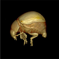
Video credit: Danny Poinapen, Joanna K. Konopka, Joseph U. Umoh, Chris J. D. Norley, Jeremy N. McNeil and David W. Holdsworth via BMC Zoology / CC BY 4.0
Leave A Comment